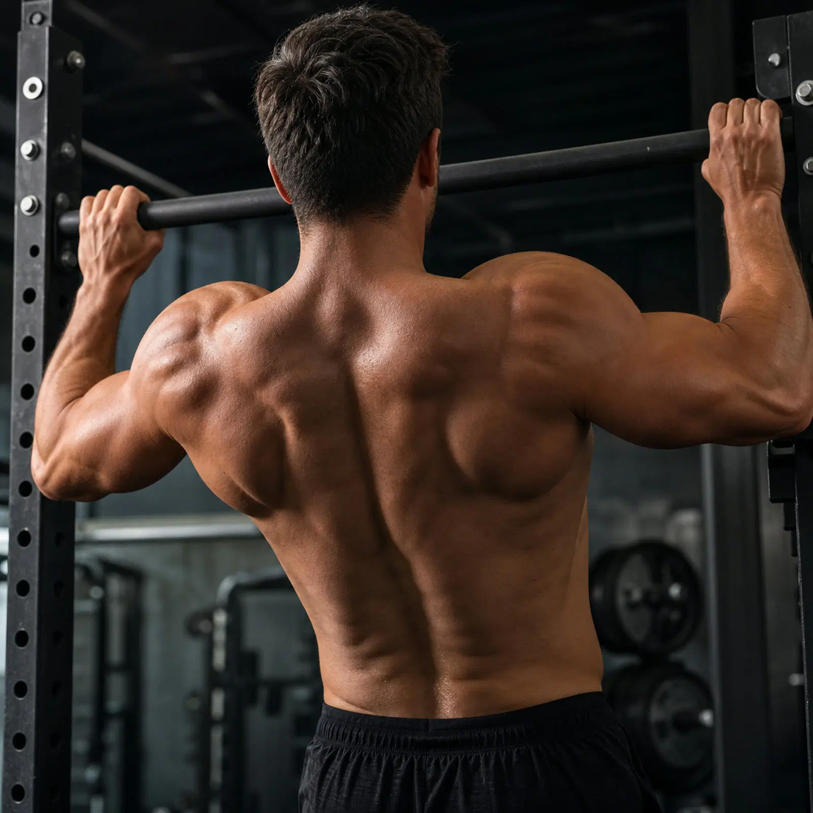
Anatomy for lifters: which muscles move which joints, so exercise choices stop being guesswork.
What do you want to do?
Start here
4 lessons · ~4 min · in this order- 1Kinesiology and Biomechanics (Introduction)Kinesiology is the science of human movement; biomechanics applies the principles of mechanics to exercise.Begin →
- 2The 12 Major Muscle GroupsFor balanced development and reduced injury risk, strengthen the 12 major muscle groups (plus obliques and adductors/abductors).1 min
- 3The Body's Three Planes of MotionThe body moves in three planes — frontal, transverse, and sagittal.1 min
- 4Muscle Action Roles (Agonist, Antagonist, Synergist)A muscle can act as a primary mover, secondary mover, antagonist, synergist/neutralizer, or stabilizer; reciprocal innervation relaxes the antagonist as the agonist contracts.1 min
Every Anatomy topic
99 topics
- Abdominals (Midsection)2 minMuscle Group · Four muscles — rectus abdominis, external oblique, internal oblique, transverse abdominis — that flex, rotate, and laterally flex the trunk and compress the abdomen.
- Abduction1 minMovement Term · Movement of a body part away from the midline (axis) of the trunk.
- Adduction1 minMovement Term · Movement of a body part toward the midline (axis) of the trunk.
- Anterior Deltoid1 minMuscle · The front deltoid head; produces forward flexion, horizontal adduction, and internal rotation — primarily a pressing muscle.
- Antigravity Musculature2 minMuscle Group · The large muscle groups that resist gravity to maintain erect posture — gastrocnemius/soleus, quadriceps, gluteus maximus, erector spinae, and abdominals.
- Atrophic Weakening Muscle Groups1 minMuscle Group · Eight major muscle groups especially prone to age-related atrophic weakening that a sound strength program should target.
- Biceps Brachii1 minMuscle · The strongest flexor of the elbow joint (long and short heads); flexes the elbow, supinates the forearm, and weakly flexes the shoulder; a 2-joint muscle (ROM 135°).
- Body Frame Types (T/A/O-Frame) — Dated Terminology, Modern Equivalent3 minConcept · A 1996 magazine series classified women into T-frame, A-frame, and O-frame body shapes with muscle-emphasis advice for visual symmetry — dated terminology now superseded by the ratio-based Inverted Triangle / Pear / Apple system, but the underlying idea (train to balance proportions) still holds.
- Body Shape — Somatotypes (Endomorph / Mesomorph / Ectomorph)3 minConcept · Sheldon's (1954) three-component somatotype system rates each person on endomorphy (fat), mesomorphy (muscle), and ectomorphy (linearity) on a 1-7 scale each — most people are a combination of two, not a single dominant type.
- Bone Tissue and Bone Health2 minStructure · Bone is living tissue that constantly remodels and adapts to mechanical load — which is why weight-bearing and resistance exercise, alongside calcium and vitamin D, build and maintain bone density.
- Bones and Levers in Movement2 minConcept · Muscles exert force on body levers (bones); the muscle's mechanical ability to exert force changes through the range of motion as lever and joint angle change.
- Brachialis1 minMuscle · The only pure flexor of the elbow joint; pulls on the ulna, which does not rotate.
- Brachioradialis1 minMuscle · A forearm flexor that flexes the elbow and both supinates and pronates the forearm.
- Calves (Gastrocnemius and Soleus)1 minMuscle Group · The calf comprises the gastrocnemius (crosses knee and ankle) and the soleus (crosses only the ankle); both plantar-flex the foot.
- Carrying Angle of the Elbow1 minStructure · Because the trochlea extends longer than the capitulum, the extended supinated forearm deviates laterally from the humerus — the carrying angle (cubital angle), ~5° in men and 10–15° in women.
- Deltoids1 minMuscle Group · The three-headed shoulder muscle — anterior, medial, and posterior deltoid — each with distinct actions.
- Depression1 minMovement Term · Downward movement returning to the normal position from elevation.
- Dorsiflexion1 minMovement Term · Movement of the top of the foot toward the anterior tibia bone.
- Elbow / Biceps Range of Motion1 minRom Reference · The biceps brachii (a 2-joint muscle) has a ROM of 135 degrees at the elbow.
- Elevation1 minMovement Term · Upward movement, as in shrugging the shoulder girdles.
- Erector Spinae (Sacrospinalis)1 minMuscle Group · Three muscle-group columns (iliocostalis, longissimus, spinalis) running skull-to-sacrum that extend, laterally flex, and rotate the vertebral column.
- Eversion1 minMovement Term · Turning the sole outward, placing weight on the inner edge of the foot.
- Extension1 minMovement Term · Straightening a joint so the bones move apart.
- External Oblique1 minMuscle · The largest, most superficial flat abdominal; flexes the trunk and rotates it toward the opposite side.
- External Rotation1 minMovement Term · Rotation with the axis of the bone turning away from the body (e.g., the humerus turned outward).
- Femur Length vs. Torso Length (Squat Mechanics)1 minConcept · Long-femured lifters lean farther forward in the squat, shifting emphasis toward the glutes and erector spinae.
- Fibularis Longus (Peroneus Longus)1 minMuscle · The lateral lower-leg muscle that everts the foot and helps support the arch — the antagonist to the tibialis posterior.
- Flexion1 minMovement Term · Bending a joint so the bones come closer together.
- Foot / Ankle Range of Motion1 minRom Reference · Plantar flexion of the foot 45°, dorsiflexion of the foot 10–15°.
- Forearm Flexors1 minMuscle Group · The forearm flexors — flexor carpi ulnaris and flexor carpi radialis — flex the wrist; trained with wrist curls.
- Gastrocnemius1 minMuscle · A 2-joint calf muscle that plantar-flexes the ankle and flexes the knee; origin at the femoral condyles, insertion at the calcaneus.
- Gluteus Maximus1 minMuscle · The strongest, largest muscle in the body; the primary hip extensor and a lateral rotator of the femur.
- Gluteus Medius1 minMuscle · A fan-shaped hip abductor under the gluteus maximus, important for balance in walking.
- Gluteus Minimus1 minMuscle · The smallest gluteal muscle; functions similarly to the gluteus medius as a hip abductor.
- Gracilis1 minMuscle · The only 2-joint hip adductor; a weak adductor and knee flexor that the body can do without.
- Hamstrings1 minMuscle Group · Three 2-joint muscles — biceps femoris, semitendinosus, semimembranosus — that flex the knee (primary) and extend the hip (secondary); knee-flexion ROM 125°.
- Hip Abductors1 minMuscle Group · The hip abductors are the gluteus medius and gluteus minimus.
- Hip Adductors1 minMuscle Group · The hip adductors are the pectineus, adductor brevis, adductor longus, adductor magnus, and gracilis.
- Hip Extensors1 minMuscle Group · The primary hip extensors are the gluteus maximus and the hamstrings.
- Hip Flexors1 minMuscle Group · The 4 major hip flexors — iliopsoas, rectus femoris, tensor fascia lata, sartorius — plus secondary flexors.
- Hip Joint Range of Motion1 minRom Reference · Hip flexion 90° (knee extended) to 120° (up to 135°, knee flexed); hyperextension 10–30°; adduction 30°; abduction 45°; medial rotation 30–45°; lateral rotation 45–60°.
- Hip Lateral Rotators (6 Short Muscles)1 minMuscle Group · Six short muscles produce lateral (outward) rotation of the hip.
- Horizontal Extension (Abduction)1 minMovement Term · Returning the humerus from the front-horizontal position back to the side-horizontal position.
- Horizontal Flexion (Adduction)1 minMovement Term · Drawing the humerus from the side-horizontal to the front-horizontal position.
- Iliopsoas1 minMuscle · The major hip flexor, made of the iliacus and psoas major; shortening it increases lumbar lordosis.
- Infraspinatus1 minMuscle · The "I" of SITS; an external rotator of the shoulder also involved in horizontal abduction and extension.
- Internal Oblique1 minMuscle · A flat abdominal sandwiched between the external obliques and transversus; its fibers run at right angles to the external oblique and it twists the trunk to the same side.
- Internal Rotation1 minMovement Term · Rotation with the axis of the bone turning toward the body (e.g., the humerus turned inward).
- Inversion (Foot Movement)1 minMovement Term · Turning the sole inward, placing weight on the outer edge of the foot.
- Joint Range of Motion Chart2 minRom Reference · Reference table of degree-of-motion values for the hip, knee, spine, arm/shoulder, neck, and foot (Kennedy & Legel, 1992).
- Kinesiology and Biomechanics (Introduction)1 minConcept · Kinesiology is the science of human movement; biomechanics applies the principles of mechanics to exercise.
- Knee Joint Range of Motion3 minRom Reference · The knee is a hinge joint that straightens to about 0° and, in healthy adults, bends to roughly 133–142° depending on age and sex (CDC reference data). The "90°" often quoted is the arc of a seated leg extension, not the knee's full range.
- Knee-Toe Relationship1 minConcept · A squat-alignment concept balancing keeping the knees from passing the toes against keeping the spine vertical — the "happy medium."
- Latissimus Dorsi2 minMuscle · A 2-joint back muscle; its strongest action is adduction (drawing the arm to the side from side-horizontal, rotating inward), with extension second.
- Levator Scapulae1 minMuscle · A neck-to-scapula strap muscle that elevates the scapula alongside the upper trapezius — the muscle most often blamed for a stiff, tight neck-shoulder junction.
- Medial Deltoid1 minMuscle · The middle deltoid head; the primary muscle of true shoulder abduction (90°), involving all three heads plus the supraspinatus.
- Multifidus3 minMuscle · A deep, segment-by-segment spinal stabilizer running from the sacrum to C2 — not a prime mover for big spinal ROM, but a key part of the local stability system that's commonly found inhibited or smaller in people with chronic low-back pain.
- Muscle Action Roles (Agonist, Antagonist, Synergist)1 minConcept · A muscle can act as a primary mover, secondary mover, antagonist, synergist/neutralizer, or stabilizer; reciprocal innervation relaxes the antagonist as the agonist contracts.
- Muscle Fiber Arrangements (Penniform vs. Fusiform)1 minConcept · The two major fiber arrangements — penniform (greater cross-section, more force) and fusiform (parallel fibers, speed but less force).
- Neck Range of Motion1 minRom Reference · Neck abduction/adduction 40°, flexion 40°, hyperextension 50°, rotation 55°.
- Pectoralis Major2 minMuscle · The chest muscle (clavicular and sternal portions); its primary action is horizontal adduction (flexion) of the arm — a powerful action.
- Pectoralis Minor1 minMuscle · A thin triangular muscle deep to the pec major that pulls the scapula forward and down — the muscle most implicated in a rounded-shoulder posture when it's chronically tight.
- Plantar Flexion1 minMovement Term · Movement of the sole downward toward the floor, as in performing calf raises.
- Posterior Deltoid1 minMuscle · The rear deltoid head; produces horizontal abduction, backward extension, and external rotation — the most under-emphasized deltoid head.
- Pronation1 minMovement Term · Rotation of the forearm that turns the palm down, rotating the radius on the ulna.
- Quadratus Lumborum1 minMuscle · A deep lower-back muscle important in lateral trunk flexion; trained with side bends and lateral pelvic tilts.
- Quadriceps2 minMuscle Group · The four knee extensors — rectus femoris, vastus medialis, vastus lateralis, vastus intermedius (rectus femoris also flexes the hip); the leg-extension arc is ~90° (knee bent to straight).
- Range of Motion (ROM)1 minConcept · The complete movement capability of a joint; train through the full ROM a joint safely allows.
- Rectus Abdominis1 minMuscle · The "abs"; a long strap-like trunk flexor split into left/right halves by the linea alba.
- Rectus Femoris1 minMuscle · A 2-joint quadriceps muscle that flexes the hip and extends the knee.
- Rhomboids (Major and Minor)1 minMuscle · Two muscles between the spine and the inner edge of the scapula that retract (draw together) and downwardly rotate the shoulder blades — key to upright posture and rowing movements.
- Rotator Cuff (SITS)1 minMuscle Group · The four rotator cuff muscles — Supraspinatus, Infraspinatus, Teres minor, Subscapularis (SITS) — that rotate and stabilize the shoulder joint.
- Sartorius1 minMuscle · The longest muscle in the body; a 2-joint hip flexor and adductor that also flexes and laterally rotates the knee.
- Scapular Movement1 minMovement Term · The scapula moves through elevation/depression, adduction (retraction)/abduction (protraction), and upward/downward rotation.
- Serratus Anterior1 minMuscle · The muscle along the rib cage that pulls the scapula forward and around the chest wall (protraction) and helps rotate it upward, acting as a stabilizer in most pressing movements.
- Shoulder / Arm Range of Motion1 minRom Reference · Arm flexion 135°, shoulder adduction 50°, shoulder hyperextension 45°, shoulder abduction 90–180°.
- Soleus1 minMuscle · A single-joint calf muscle that plantar-flexes the ankle; trained with seated calf raises.
- Spinal Column (Vertebral Column)2 minStructure · A column of 33 vertebrae and 23 intervertebral discs with 4 curves that supports the trunk, protects the spinal cord, and absorbs shock.
- Spine Range of Motion1 minRom Reference · Spinal lateral bending 30°, rotation 40°, flexion 70° (standing and lying), hyperextension 70°.
- Sternocleidomastoid1 minMuscle · A neck muscle and the first of the atrophic-weakening muscle groups; trained with neck machines / neck strap with weight plate.
- Subscapularis1 minMuscle · The "S" of SITS; the internal rotator of the shoulder, also involved in extension and adduction.
- Supination1 minMovement Term · Rotation of the forearm that turns the palm up, rotating the radius on the ulna.
- Supraspinatus1 minMuscle · The "S" of SITS; it abducts the humerus and does not rotate it.
- Tensor Fascia Lata1 minMuscle · A hip flexor that also abducts and medially rotates the femur, inserting via the iliotibial (IT) band.
- Teres Major1 minMuscle · The "lat's little helper" — a scapula-to-humerus synergist that extends, adducts, and internally rotates the shoulder alongside the latissimus dorsi.
- Teres Minor1 minMuscle · The "T" of SITS; an external rotator of the shoulder also involved in horizontal abduction and extension.
- The 12 Major Muscle Groups1 minMuscle Group · For balanced development and reduced injury risk, strengthen the 12 major muscle groups (plus obliques and adductors/abductors).
- The Body's Three Planes of Motion1 minConcept · The body moves in three planes — frontal, transverse, and sagittal.
- The Hip Complex1 minStructure · The musculature spanning the hip — including the iliopsoas, tensor fasciae latae, sartorius and the iliotibial band — that flexes, extends, adducts, abducts, and rotates the hip.
- Tibialis Anterior1 minMuscle · The shin muscle that dorsiflexes the foot (lifts the top of the foot toward the shin) — the antagonist to the calf muscles.
- Tibialis Posterior1 minMuscle · The deep calf muscle that inverts the foot and is the primary dynamic support of the medial arch — the antagonist to the fibularis (peroneal) muscles.
- Transverse Abdominis1 minMuscle · The deepest abdominal muscle; compresses the abdomen, supports the abdominal contents, and aids lower-back stability.
- Trapezius1 minMuscle · A muscle that functions as three different muscles — upper, middle, and lower traps — moving the scapula.
- Triceps Brachii1 minMuscle · The three-headed elbow extensor (long, medial, lateral heads); the long head also assists shoulder extension.
- Two-Joint (2-Joint) Muscles1 minConcept · Muscles crossing two joints generate the greatest force when not required to shorten over both joints simultaneously; stabilize one joint when training them.
- Vastus Intermedius2 minMuscle · The deepest of the four quadriceps heads, sitting directly beneath rectus femoris where it can't be seen or palpated separately — a pure knee extensor that still moves real weight even though it's functionally invisible.
- Vastus Lateralis2 minMuscle · The largest and outermost of the four quadriceps heads — a pure knee extensor running down the lateral thigh, and the muscle most often biopsied in exercise-physiology research because of its size and easy access.
- Vastus Medialis2 minMuscle · The medial quadriceps head, whose distal fibers (the "VMO") angle sharply inward and fire hardest in the last 15° of knee extension — the medial counterbalance that keeps the kneecap tracking straight.
- Wrist Extensors1 minMuscle Group · The extensor carpi radialis longus/brevis and extensor carpi ulnaris — the mirror-image group to the wrist flexors, trained with reverse curls and reverse wrist curls.
Explore connections
How Anatomy links to other categories — line thickness is link density in the graph, not an editorial rating.